
撰文|路玉蝶(2025级博士研究生)
审核|Mohamed Elsherif Badr
Academic Presentation: Basic Information
Time and Venue: Wednesday June 24, 2026, 4:00 P.M., Innovation Hub, 2nd Floor, North Basic Research Building
Speaker: Linrong Lu, Professor, Zhejiang University
Lab focus: T-cell development, differentiation, and effector function regulation

Academic Presentation: Main Content
· Thymocyte selection: The role of Tespa1
· Th differentiation: MINK1/PP2A axis
· CD8 regulatory T cells: Time to revisit
Thymocyte selection: The role of Tespa1
Background and Significance
Research Background
Thymic Positive Selection: A Critical Checkpoint in T Cell Development
T cells undergo development within the thymus, with positive selection serving as the central checkpoint. During this process, double-positive (DP) thymocytes must express T cell receptors (TCRs) capable of recognizing self-peptide–MHC complexes to receive survival signals and differentiate into mature CD4⁺ or CD8⁺ single-positive (SP) T cells. Those failing to engage ligands with sufficient affinity undergo apoptosis—a process termed "death by neglect."
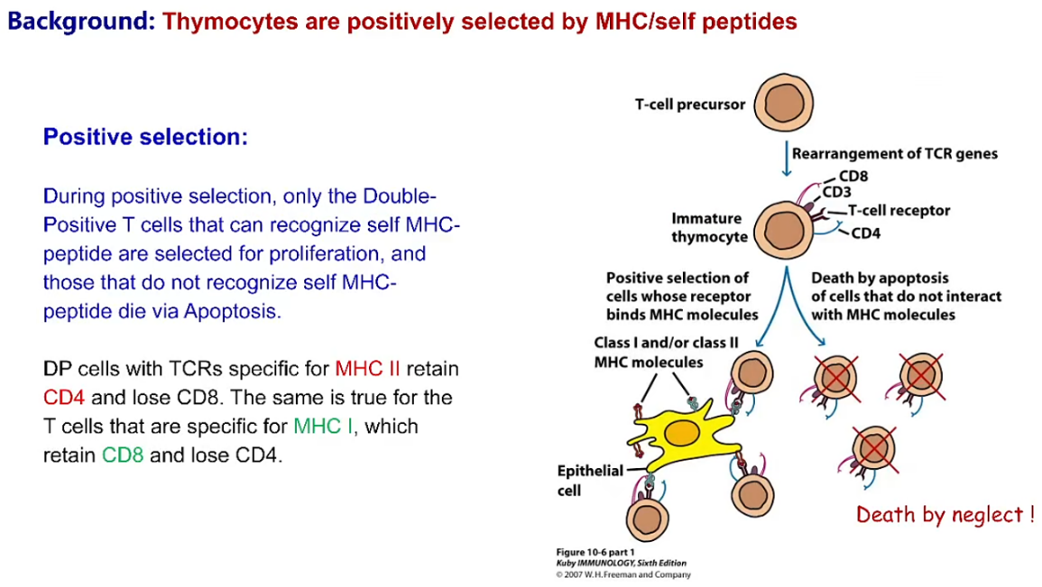
This process presents a long-standing paradox: DP thymocytes express lower surface TCR levels than mature T cells, and the ligands that drive positive selection are low-affinity self-peptide–MHC complexes. Yet DP cells respond robustly to these weak signals, whereas mature T cells require high-affinity antigens for activation.
Core Question: How do DP thymocytes achieve high sensitivity to weak ligands despite low TCR density? It has been postulated that thymus-specific signal-enhancing mechanisms exist, but definitive molecular explanations have long remained elusive.
Discovery of Tespa1
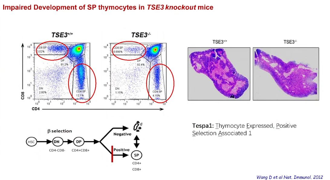
In 2012, Professor Lu's team identified a novel gene highly expressed in DP thymocytes through transcriptomic profiling across distinct thymic developmental stages, naming it Tespa1 (thymocyte-expressed, positive selection-associated 1). Tespa1-deficient mice exhibit a classic positive selection defect—marked reductions in both CD4⁺ and CD8⁺ SP thymocytes. However, the molecular mechanism by which Tespa1 regulates TCR signaling remained unknown.
Key Scientific Questions
Which molecules does Tespa1 interact with within the TCR signalosome?
By what mechanism does Tespa1 enhance TCR signaling?
Why is Tespa1 functionally required only in DP thymocytes but dispensable in mature T cells?
Significance
Answer a key remaining question in TCR signaling during thymocyte selection-How do DP thymocytes coordinate low TCR expression and cross-reactive low affinity ligands with high signaling sensitivity?
Research Rationale and Significance
Gene Discovery – Tespa1
To explore the function of Tespa1, the team generated Tespa1 knockout mice. Phenotypic analysis revealed a critical finding: Tespa1 deficiency resulted in a blockade of thymocyte transition from the DP to SP stage, severely impaired positive selection, and markedly reduced peripheral CD4⁺ and CD8⁺ T cell numbers.
Mechanistic Study – How Tespa1 Regulates TCR Signaling
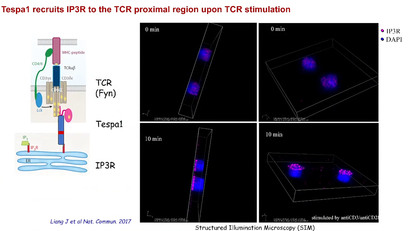
Interactome Screening: A Flag-Tespa1 Jurkat stable cell line was established. Following anti-CD3/CD28 stimulation, anti-Flag pull-down coupled with mass spectrometry was performed to identify Tespa1-interacting proteins. In addition to the known interactors PLCγ1 and Grb2, Tespa1 was found to interact with IP3R1, IP3R2, and IP3R3—calcium channel proteins on the ER membrane—for the first time.
So why signaling protein binds to Ino channel in the ER? They found that determining the translocation of IP3R on the ER to the signal complex of TCR proximal through structured illumination microscopy. All the IP3R binds Tespa1 and closed to the region of TCR proximal.
Thymic Developmental Specificity
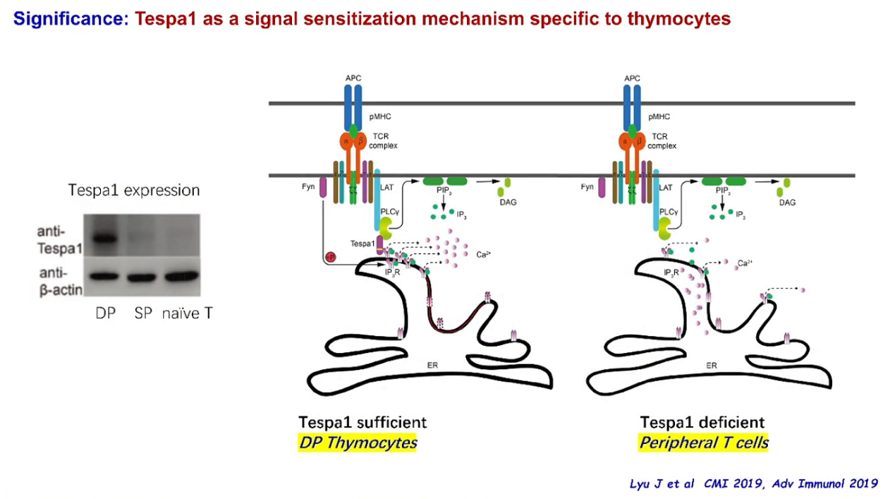
Expression Characteristics: Tespa1 is highly expressed in DP thymocytes but significantly downregulated in SP thymocytes and peripheral T cells.
Conditional Knockout Validation: Following Tespa1 deletion in peripheral T cells, IP3R cannot reach to the region of TCR complex.
Key Findings and Conclusions
Key Findings:
1. Tespa1 is a novel component of the TCR signalosome and plays a critical role in regulating TCR signaling during T cell selection and maturation.
2. Tespa1 recruits IP3R1 to the TCR-proximal region upon TCR stimulation.
3. Tespa1 functions as a signal sensitization mechanism specific to thymocytes.
Conclusions:
By physically recruiting IP3R1 to the TCR-proximal membrane region, Tespa1 achieves an order-of-magnitude enhancement in signaling efficiency, thereby ensuring that DP thymocytes can respond to low-affinity ligands despite low TCR density and successfully complete positive selection.
Th differentiation: MINK1/PP2A axis
Background and Significance
Research Background
It is well established that CD4⁺ T cells can differentiate into distinct helper subsets, with Th1 and Th2 being identified in the 1980s. Subsequently, the CD4⁺ T-helper cell family has been expanded to include several other subsets, such as Th1, Th2, Th17, Treg, and Tfh. Among these, hyperactivation of the Th17 subset is closely associated with the pathogenesis of multiple autoimmune diseases. The present study focuses on the role of kinases in regulating Th17 differentiation.
Significance
The first work discover MINK1 restrains Th17 differentiation through direct phosphorylation of SMAD2. Also they find PP2A promotes Th17 differentiation through the same pathway.
Research Rationale and Significance
MINK1 Study
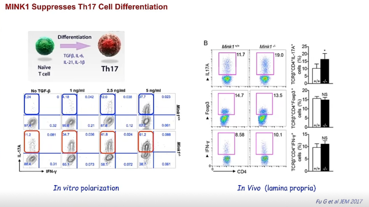
MINK1 Knockout phenotype: Th17 accumulation in vivo.
In vitro differentiation: When they stimulate Naïve T cells with different TGFβ concentration. And they found that MINK1 specifically suppresses Th17.
Experimental autoimmune encephalomyelitis (EAE) model: When they knockout MINK1 in mouse, they find mouse have more Th17 dependent disease EAE.
PP2A Study
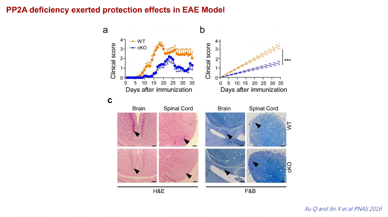
Since MINK1 co-localizes with PP2A within the STRIPAK complex, they sought to determine whether PP2A is also involved in Th17 differentiation. T-cell-specific PP2A knockout mice were generated, and in these mice, Th17 differentiation was reduced to about 50% of wild-type levels. Furthermore, PP2A deficiency provided protection against EAE, suggesting a critical role for PP2A in Th17-driven autoimmune pathogenesis.
Key Findings and Conclusions
1. MINK1 acts as a negative regulator of Th17 differentiation.
2. PP2A functions as a positive regulator of Th17 differentiation.
CD8 Regulatory T Cells: Time to Revisit
Research Background
One of the central challenges in immunology is understanding how the immune system suppresses excessive immune responses without compromising protective immunity. While the biology of CD4 regulatory T (Treg) cells has been extensively studied, the mechanisms underlying CD8 regulatory T (CD8 Treg) cell function remain poorly understood. Although CD8 Tregs were identified decades before CD4 Tregs, progress in the field has been hindered by the absence of a lineage-defining transcription factor and the lack of reliable surface markers. Consequently, the identity, antigen specificity, and physiological function of CD8 Tregs have remained controversial.
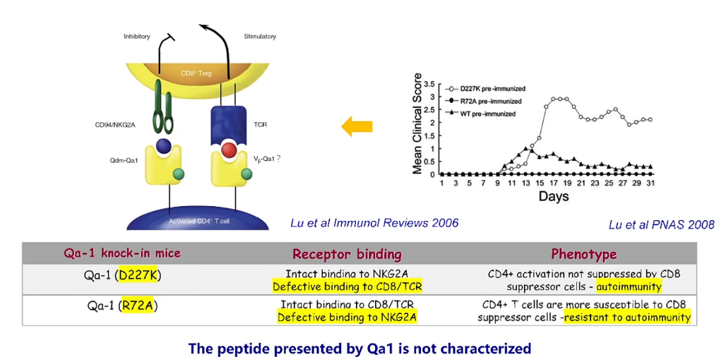
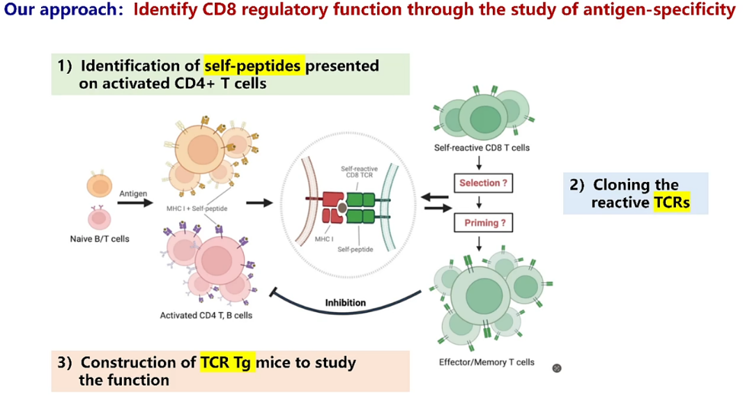
This seminar addressed these longstanding questions by investigating whether CD8 Tregs recognize activated CD4 T cells in an antigen-specific manner. The work was based on previous studies demonstrating that the non-classical MHC class Ib molecule Qa-1 mediates communication between activated CD4 T cells and CD8 Tregs. Mutational analyses have shown that disrupting TCR recognition, on CD8 Teg, of Qa-1, on CD4 T cells, abolishes CD8-mediated suppression, whereas preserving TCR recognition enhances protection from autoimmune disease. Together with evidence that classical MHC class I molecules can also participate in CD8 Treg recognition, these findings led to the hypothesis that activated CD4 T cells present endogenous self-peptides that are specifically recognized by regulatory CD8 T cells.
Approach:
Research Rationale and Key Findings
To test this hypothesis, the investigators first examined whether activated CD4 T cells express the machinery required for antigen presentation through classical MHC class I molecules. Both in vitro and in vivo activation experiments demonstrated marked upregulation of classical MHCI molecule, H2-Kb and H2-Db, on activated CD4 T cells. RNA sequencing further revealed coordinated induction of genes involved in antigen processing and presentation, and these transcriptional changes were confirmed at the protein level by Western blotting. These results established that activated CD4 T cells are capable of efficiently presenting endogenous peptides through MHC class I.
The next objective was to identify the endogenous peptides presented by activated CD4 T cells. Activated CD4 T cells were isolated, MHC class I molecules were immunoprecipitated, and bound peptides were analyzed by mass spectrometry. This approach identified nine endogenous self-peptides. Their ability to bind MHC class I molecules was subsequently evaluated using the TAP-deficient RMA-S cell line, which increase the expression level of MHCI on their surfaces in the presence of exogenous peptides capable of binding to MHCI. Most peptides enhanced surface MHC class I expression in a concentration-dependent manner, confirming that they form stable peptide-MHC complexes suitable for T-cell recognition.
The investigators then determined whether these self-peptides could elicit functional CD8 T-cell responses. Mice were immunized with each peptide in complete Freund's adjuvant, and peptide-specific responses were evaluated by intracellular cytokine staining and IFN-γ ELISPOT assays. Most peptides induced robust antigen-specific CD8 T-cell responses. More importantly, immunization with four selected peptides completely protected mice from experimental autoimmune encephalomyelitis (EAE). This protection was lost following CD8 depletion, demonstrating that CD8 T cells were essential for disease suppression. Furthermore, adoptive transfer of peptide-primed CD8 T cells into naïve recipients conferred protection against EAE, establishing that these cells were sufficient to mediate immune regulation.
To define the antigen receptors responsible for this activity, mice were immunized with the protective peptide mixture, responding CD8 T cells were isolated, and single-cell TCR sequencing was performed. Several antigen-reactive TCRs were identified, with one clone (TCR4) exhibiting the strongest response. Further analyses demonstrated that TCR4 specifically recognized the peptide pDYHC1. Remarkably, immunization with pDYHC1 alone reproduced the protective effects observed with the four-peptide mixture, indicating that a single endogenous peptide could generate protective regulatory CD8 T-cell responses.
The investigators next generated TCR4 transgenic mice to characterize this regulatory population in greater detail. Transcriptomic analysis revealed that these cells did not constitutively express Ly49, a previously proposed CD8 Treg marker. However, they displayed a gene-expression program closely resembling previously described Ly49⁺ regulatory CD8 T cells and were enriched for characteristics associated with self-reactive CD44⁺CD122⁺ memory-like CD8 T cells. Although these cells exhibited features of functional anergy in vitro, they could be efficiently activated both in vitro upon stimulating them in the presence of co-stimulation and in vivo. Upon activation, TCR4 CD8 T cells proliferated vigorously, produced IFN-γ, exhibited antigen-specific cytotoxicity, suppressed pathogenic CD4 T-cell responses, and protected recipient mice from multiple autoimmune diseases, including EAE, transfer colitis, and experimental autoimmune uveitis. These findings provide compelling evidence that antigen-specific, self-reactive CD8 T cells can function as regulatory T cells following appropriate activation.
Q&A Highlights
1. Q: For different autoimmune diseases, do you think they have same or different CD8 regulatory T cells?
A: So this is a basic question about general antigen specificity. We do believe this mechanism is general because we isolate these peptides from in vitro activated CD4 T cells and could work on different disease models.
2. Q: Have you ever tried human T cells to isolate CD8 regulatory T cells?
A: We haven't finished yet, but we do have peptides from human activated CD4 T cells. And we knew they can be presented as well as if we take human blood CD8 T cells and some of them can stimulate IFNg expression.
3. Q: As you mentioned, in CD4 Treg cells, they have Foxp3 and CD25 as markers but CD8 regulatory T cells don't have clear markers whatever surface marker or TFs. So what is the special features of these identified CD8 regulatory T cells?
A: You may never be able to identify using surface markers. So the significant finding is that if we do find dominant features for example, antigen for that and you can use tetramer to enrich them. In mouse, we transfer our TCR4 cells into host mouse with different markers. So we can then trace these cells to see whether in the certain stage or different condition they can show some special characteristics.
4. Q: CD4 regulatory T cells can be induce in vitro with IL-2 and TGFβ,so can CD8 regulatory T cells be induced in vitro?
A: If we find antigen specificity, it is easier to induce in vitro.
Q: Can they be induced from naïve cells?
A: Possibly not, because CD8 T cells need to be primed and I think priming only can be done in vivo.
5. Q: How can distinguish CD8 autoreactive T cells and CD8 regulatory T cells?
A: We can use peptide-MHC-tetramer to satin them.
Q: But you cannot list all antigens
A: Of course, it depends on the purpose. If you want to character their function. That is enough.
6. Q: You identified those self-peptides, they only come from activated CD4 T cells or CD4 cross-present peptides from other cells?
A: We don't think CD4 can cross-present peptides. All the souse protein of these peptides are expressed in the activated CD4 T cells. We all believe that they come from the endogenous protein of CD4.
Q: So they are limited in CD4
A: Yes, but the question is other cells also express these source proteins.
7. Q: Do you think what percentage of so called CD8 regulatory T cells in normal or EAE model?
A:In EAE model, we use tetramer to stain antigen-specific CD8, we see very few even cannot see it. However, if you isolate CD8 regulatory T cells from mouse and restimulated in vitro, we can see reasonable percentage.
Learning Takeaways and Reflections
Research Approaches and Skills I Learned
This seminar showcased a rigorous hypothesis-driven pipeline that integrates molecular, cellular, and in vivo approaches. I learned how to combine RNA-seq and Western blotting to confirm gene expression changes, use immunoprecipitation coupled with mass spectrometry to identify endogenous peptide ligands, and employ the TAP-deficient RMA-S cell line to validate peptide–MHC binding efficiently. Functionally, the study elegantly employed disease models, cell depletion, and adoptive transfer to prove both necessity and sufficiency of the regulatory cells. The use of single-cell TCR sequencing and subsequent generation of a transgenic mouse model was particularly instructive, demonstrating how to isolate a key functional clone from a polyclonal response and characterize its biology at the molecular level.
Implications for My Own Research
This work has fundamentally broadened my perspective, compelling me to actively consider CD8 regulatory T cells—not just CD4 Tregs—in my autoimmune disease models. The finding that a single self-peptide and its cognate TCR can confer protection highlights the importance of mapping the self-peptide repertoire presented in inflamed tissues, rather than relying solely on broad phenotypic markers. More importantly, the study provides a clear experimental roadmap that I can directly follow: from antigen identification and specificity validation, through functional testing in vivo, to pinpointing the critical TCR and creating a transgenic system for mechanistic dissection. This stepwise strategy will be invaluable for translating observational findings into actionable therapeutic targets in my own research.


 北京市丰台区右安门外西头条10号
 010-86738999
 cimr@cimrbj.ac.cn


 En
En

